
Liver on LabChip
|
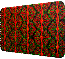
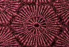
A laser diffraction-induced dielectrophoresis
(DEP) phenomenon for the patterning and manipulation of individual HepG2
cells and polystyrene beads via positive/negative DEP forces is developed.
The optoelectronic substrate was fabricated by organic photoconductive
material, TiOPc, via the spin-coating process on
the indium tin oxide glass surface. A piece of square aperture array grid
grating was utilized to transform the collimating He-Ne laser beam into the
multi-spot diffraction pattern which forms the virtual electrodes as the TiOPc-caoting surface was illuminated by the multi-spot
diffraction light pattern. HepG2 cells were trapped at the spot centers and
polystyrene beads were trapped within the dim region of the illuminating
image. The Fresnel diffraction image illustrated the distribution of trapped microparticles. This concept of utilizing laser
diffraction image to generate virtual electrodes on our TiOPc-based
optoelectronic DEP chip extends the applications of optoelectronic dielectrophoretic manipulation.
|
|
Demonstration (by Dr. Chen-Ta Ho at Micro-Systems
and Control Lab.) |
|
|
|
|
|
Selected Publication |
|
[1] Chen-Ta Ho, Ruei-Zeng Lin, Rong-Jhe Chen, Chung-Kuang
Chin, Song-En Gong, Hwan-You Chang, Hwei-Ling Peng, Long Hsu, Tri-Rung Yew, Shau-Feng Chang and Cheng-Hsien Liu, “Liver-cell
patterning Lab Chip: mimicking the morphology of liver lobule tissue,” Lab on Chip, 2013, 13, 3578-3587 (21
September 2013, Issue 18) [2] Chen-Ta
Ho , Ruei-Zeng Lin, Wen-Yu Chang, Hwan-You Chang ,
Cheng-Hsien Liu, “Rapid heterogeneous liver-cell on-chip patterning via
the enhanced field-induced dielectrophoresis
trap,” Lab on a Chip, 2006,
6, 724 – 734 |